Okay, let’s dive into some interesting medical imagery! I recently came across some images that I thought were really fascinating and worth sharing. They offer a glimpse into the intricate details of the human body, highlighting both normal anatomy and the tools we use to understand it better.
Diaphragm Impressions on the Liver

This image shows something that might seem a little strange at first glance: indentations on the surface of the liver. These are actually completely normal and are caused by the diaphragm pressing against the liver. The diaphragm, a large muscle located at the base of the chest cavity, plays a crucial role in breathing. As we inhale and exhale, the diaphragm moves up and down, and its proximity to the liver can create these visible impressions. It’s a perfect illustration of how closely connected our internal organs are and how their movements can affect each other. Doctors are well aware of these indentations and recognize them as a normal anatomical feature, so there’s no cause for alarm if you ever see them on a medical scan. They are often pointed out in medical training to avoid misinterpreting them as something pathological.
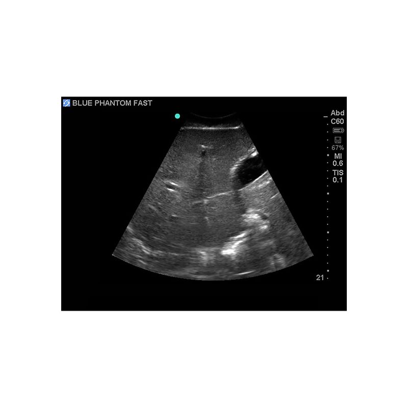
FAST Exam Ultrasound Training Model
This image showcases a valuable tool in medical education: an ultrasound training model. The “FAST” exam, which stands for Focused Assessment with Sonography for Trauma, is a rapid ultrasound examination performed on trauma patients to quickly identify signs of internal bleeding. In emergency situations, time is of the essence, and the FAST exam allows medical professionals to make critical decisions quickly and accurately. However, mastering the technique requires practice, and that’s where these training models come in. They provide a realistic simulation of the human body, allowing students and medical professionals to hone their ultrasound skills in a safe and controlled environment. They can practice identifying different anatomical structures, recognizing signs of fluid accumulation (which could indicate internal bleeding), and perfecting their scanning techniques. The use of these models significantly improves the accuracy and efficiency of FAST exams, ultimately leading to better patient outcomes in trauma situations. The details within the model, like the varying tissue densities and simulated fluid pockets, allow for a very realistic training experience, ensuring that those learning the technique are well-prepared for real-world scenarios. These models are invaluable for continual learning and improvement in emergency medicine.
So, there you have it – a glimpse into both the fascinating normal anatomy of the liver and the tools used to train medical professionals in life-saving techniques. From diaphragm indentations to ultrasound training models, the world of medicine is constantly evolving, offering us new insights and better ways to care for our health.
If you are searching about 2 A standard longitudinal section of the liver. The diaphragm (superior you’ve visit to the right place. We have 10 Pictures about 2 A standard longitudinal section of the liver. The diaphragm (superior like liver, diaphragm, vessels Diagram | Quizlet, Liver diaphragm | LPCLPC and also Diaphragmatic-hepatic window showing liver (L), diaphragm (D) and large. Here it is:
2 A Standard Longitudinal Section Of The Liver. The Diaphragm (superior

www.researchgate.net
Diaphragm Impressions On The Liver-These Are Normal Findings (white

www.researchgate.net
FAST Exam Ultrasound Training Model
medicalskillstrainers.cae.com
Liver Diaphragm | LPCLPC

liverandpancreasclinic.com
liver diaphragm hemangioma hepatocellular
Diaphragmatic-hepatic Window Showing Liver (L), Diaphragm (D) And Large

www.researchgate.net
FAST Exam Ultrasound Training Model
medicalskillstrainers.cae.com
Liver, Diaphragm, Vessels Diagram | Quizlet

quizlet.com
Liver Exam 1 Review Flashcards | Quizlet

quizlet.com
FAST Exam Ultrasound Training Model
medicalskillstrainers.cae.com
Solved The Liver Is Proximal To The Diaphragm.True Or False | Chegg.com
www.chegg.com
Diaphragm impressions on the liver-these are normal findings (white. Liver exam 1 review flashcards. Liver diaphragm


:max_bytes(150000):strip_icc()/008_how-to-factory-reset-a-lenovo-laptop-5115817-a67348722ce94f9783881ea29e596310.jpg)